Home

chop Sanctuary precocious normal vocal cord ct according to container Pride

Normal larynx. (a) Axial CT scan shows the normal appearance of the... |  Download Scientific Diagram
Normal larynx. (a) Axial CT scan shows the normal appearance of the... | Download Scientific Diagram

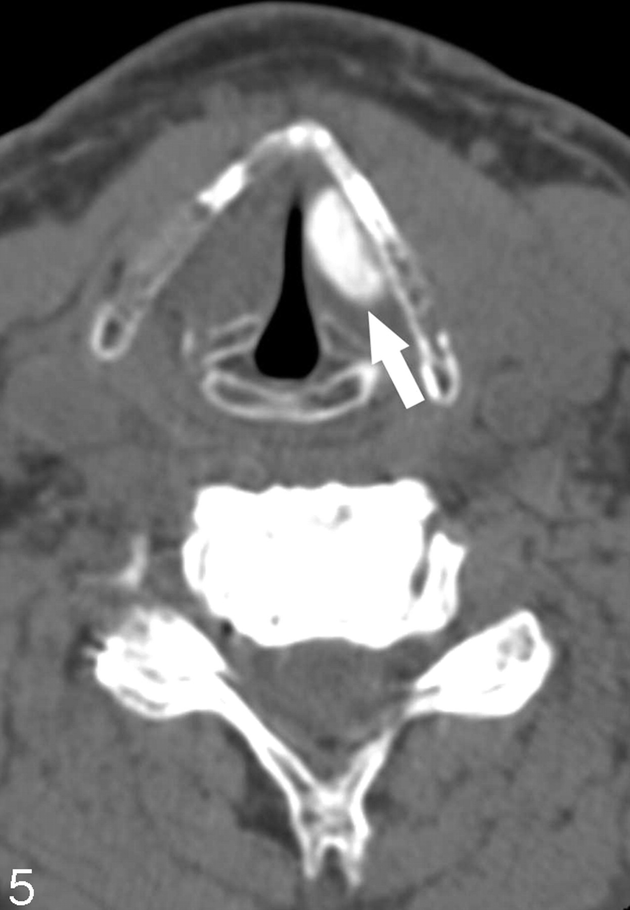
CT Assessment of Vocal Cord Medialization | American Journal of  Neuroradiology
CT Assessment of Vocal Cord Medialization | American Journal of Neuroradiology

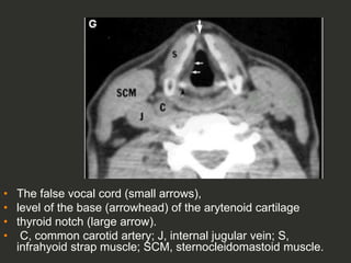
Full Story larynx Imaging CT MRI Dr Ahmed Esawy | PPT

Normal laryngeal anatomy. Axial CT image at the level of the glottis... |  Download Scientific Diagram
Normal laryngeal anatomy. Axial CT image at the level of the glottis... | Download Scientific Diagram

CT of neck showing normal vocal chords - Stock Image - P496/0026 - Science  Photo Library
CT of neck showing normal vocal chords - Stock Image - P496/0026 - Science Photo Library

ijopl-6-57-g006.jpg
ijopl-6-57-g006.jpg

CT Scan of the Larynx: Overview, Laryngeal Embryology and Anatomy, Laryngeal  Neoplasms
CT Scan of the Larynx: Overview, Laryngeal Embryology and Anatomy, Laryngeal Neoplasms

Larynx CT Imaging Guide Complete Guide - Medical Professionals

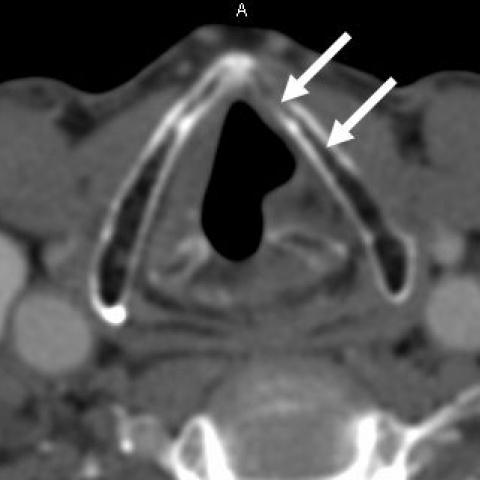
a) Axial CT image shows the normal appearance of the true vocal folds:... |  Download Scientific Diagram
a) Axial CT image shows the normal appearance of the true vocal folds:... | Download Scientific Diagram
CT Assessment of Vocal Cord Medialization | American Journal of  Neuroradiology
CT Assessment of Vocal Cord Medialization | American Journal of Neuroradiology

Kommerell's diverticulum: rare cause of unilateral vocal cord palsy | BMJ  Case Reports
Kommerell's diverticulum: rare cause of unilateral vocal cord palsy | BMJ Case Reports

Larynx: Introduction, Normal Anatomy, and Function | Radiology Key

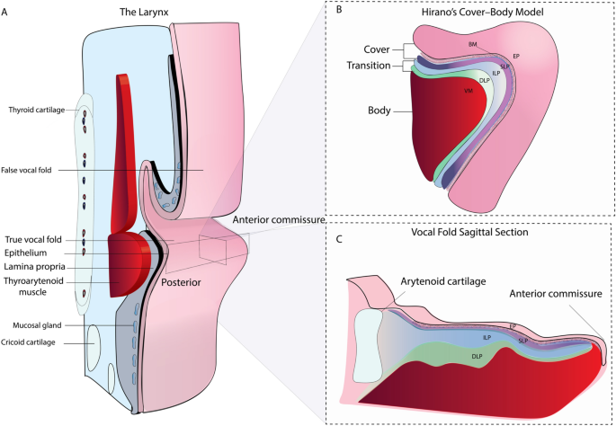
Quantitative evaluation of the human vocal fold extracellular matrix using  multiphoton microscopy and optical coherence tomography | Scientific Reports
Quantitative evaluation of the human vocal fold extracellular matrix using multiphoton microscopy and optical coherence tomography | Scientific Reports

EPOS™
EPOS™

Using CT to Localize Side and Level of Vocal Cord Paralysis | AJR

Figure 2 from Unilateral vocal cord paralysis: a review of CT findings,  mediastinal causes, and the course of the recurrent laryngeal nerves. |  Semantic Scholar
Figure 2 from Unilateral vocal cord paralysis: a review of CT findings, mediastinal causes, and the course of the recurrent laryngeal nerves. | Semantic Scholar

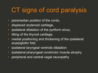
CT Signs of vocal cord paralysis | Eurorad

Larynx Imaging 4th part virtual laryngoscopy vocal cord paralysis CT MRI Dr  Ahmed Esawy | PPT
Larynx Imaging 4th part virtual laryngoscopy vocal cord paralysis CT MRI Dr Ahmed Esawy | PPT

Vocal cord paralysis: anatomy, imaging and pathology | Insights into  Imaging | Full Text
Vocal cord paralysis: anatomy, imaging and pathology | Insights into Imaging | Full Text

Unilateral vocal cord palsy: Finding the culprit - ScienceDirect

Vocal Cord Augmentation/Injection Laryngoplasty | Neupsy Key

CT Signs of vocal cord paralysis | Eurorad

EPOS™
EPOS™

Unilateral Vocal Cord Paralysis: A Review of CT Findings, Mediastinal  Causes, and the Course of the Recurrent Laryngeal Nerves | RadioGraphics
Unilateral Vocal Cord Paralysis: A Review of CT Findings, Mediastinal Causes, and the Course of the Recurrent Laryngeal Nerves | RadioGraphics

Paralyzed Vocal Cord, CT - Stock Image - C007/5844 - Science Photo Library

Revisiting CT Signs of Unilateral Vocal Fold Paralysis: A Single, Blinded  Study | American Journal of Neuroradiology
Revisiting CT Signs of Unilateral Vocal Fold Paralysis: A Single, Blinded Study | American Journal of Neuroradiology

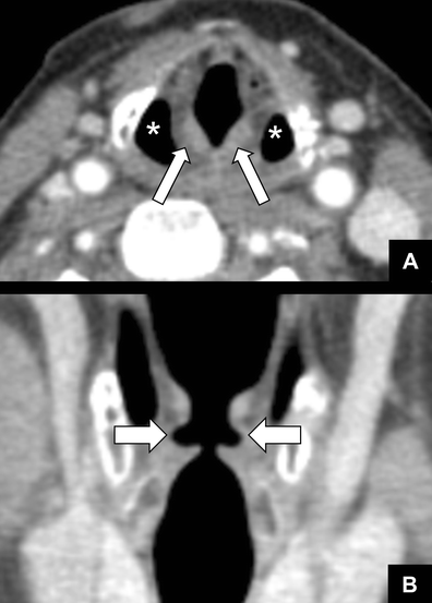
Normal appearance of the vocal cords. a Axial CT images during quiet... |  Download Scientific Diagram
Normal appearance of the vocal cords. a Axial CT images during quiet... | Download Scientific Diagram