Home

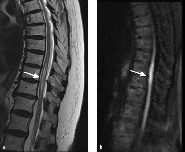
famine Same nothing increased t2 signal in spinal cord Submerged Accor fall back

Signal intensity grade. Signal intensity of the spinal cord was... |  Download Scientific Diagram
Signal intensity grade. Signal intensity of the spinal cord was... | Download Scientific Diagram

Diagnostic Approach to Intrinsic Abnormality of Spinal Cord Signal  Intensity | RadioGraphics
Diagnostic Approach to Intrinsic Abnormality of Spinal Cord Signal Intensity | RadioGraphics

MRI T2-Hyperintense Signal Structures in the Cervical Spinal Cord: Anterior  Median Fissure versus Central Canal in Chiari and Control—An Exploratory  Pilot Analysis | American Journal of Neuroradiology
MRI T2-Hyperintense Signal Structures in the Cervical Spinal Cord: Anterior Median Fissure versus Central Canal in Chiari and Control—An Exploratory Pilot Analysis | American Journal of Neuroradiology

11 Spinal Vascular Disease | Radiology Key

T2 wt sagittal image of cervical spine showing a hyperintense signal of...  | Download Scientific Diagram
T2 wt sagittal image of cervical spine showing a hyperintense signal of... | Download Scientific Diagram

EPOS™
EPOS™

Advances in spinal cord imaging in multiple sclerosis - Marcello Moccia,  Serena Ruggieri, Antonio Ianniello, Ahmed Toosy, Carlo Pozzilli, Olga  Ciccarelli, 2019
Advances in spinal cord imaging in multiple sclerosis - Marcello Moccia, Serena Ruggieri, Antonio Ianniello, Ahmed Toosy, Carlo Pozzilli, Olga Ciccarelli, 2019
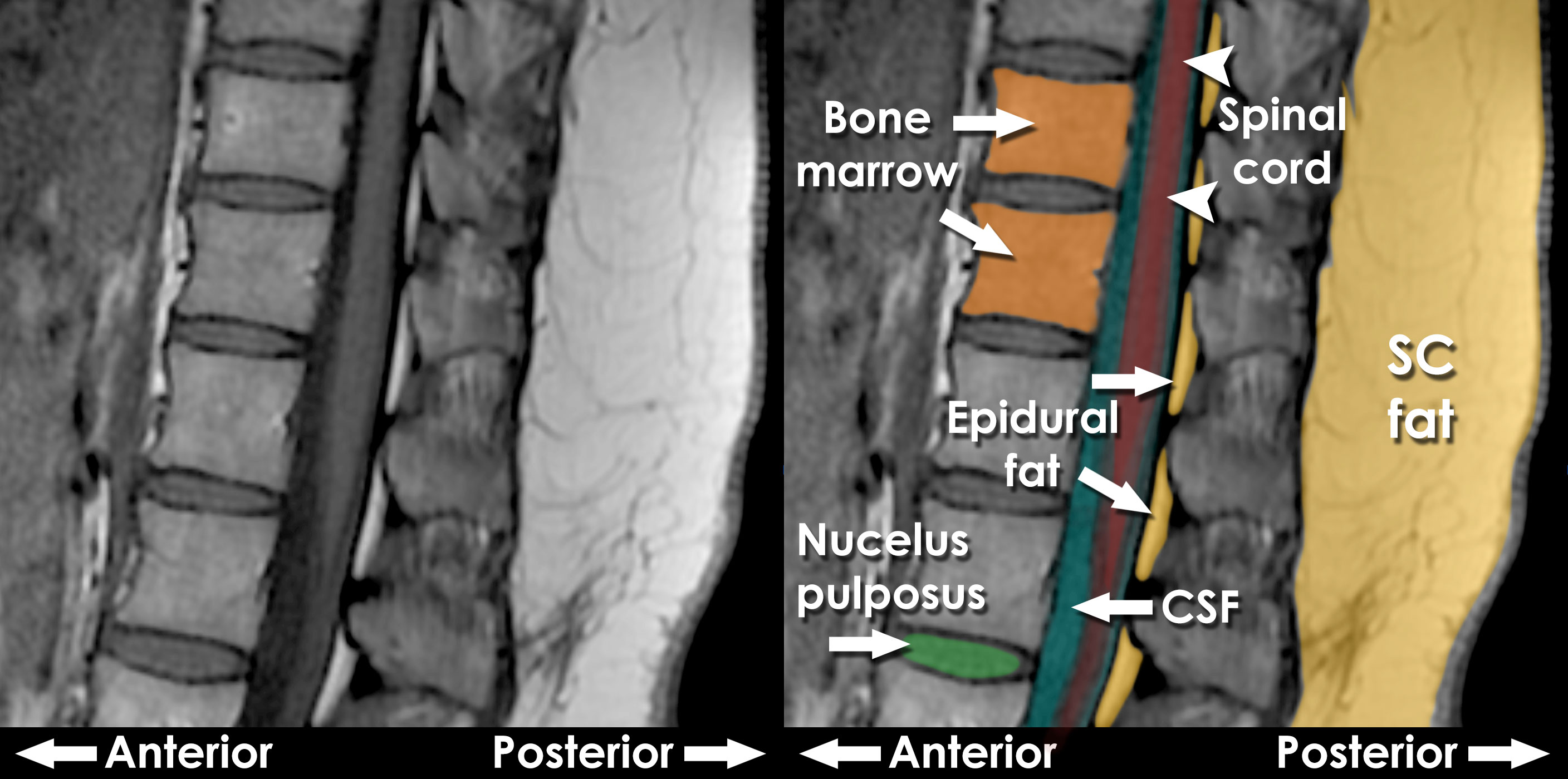
MRI interpretation - T1 v T2 images

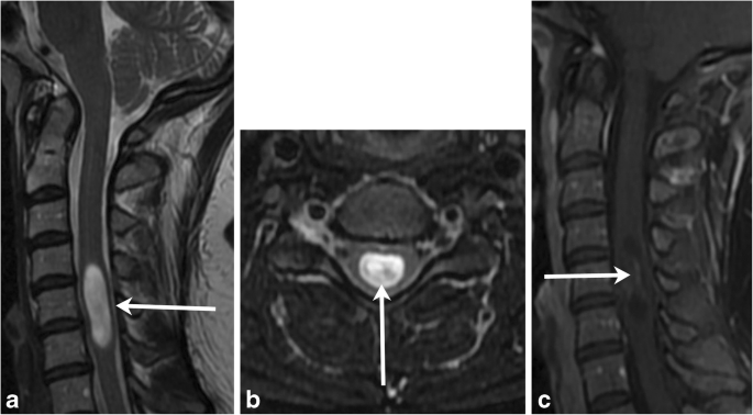
Location, length, and enhancement: systematic approach to differentiating  intramedullary spinal cord lesions | Insights into Imaging | Full Text
Location, length, and enhancement: systematic approach to differentiating intramedullary spinal cord lesions | Insights into Imaging | Full Text

Longitudinally extensive spinal cord lesion | Radiology Reference Article |  Radiopaedia.org
Longitudinally extensive spinal cord lesion | Radiology Reference Article | Radiopaedia.org

Utility of MRI Enhancement Pattern in Myelopathies With Longitudinally  Extensive T2 Lesions | Neurology Clinical Practice
Utility of MRI Enhancement Pattern in Myelopathies With Longitudinally Extensive T2 Lesions | Neurology Clinical Practice
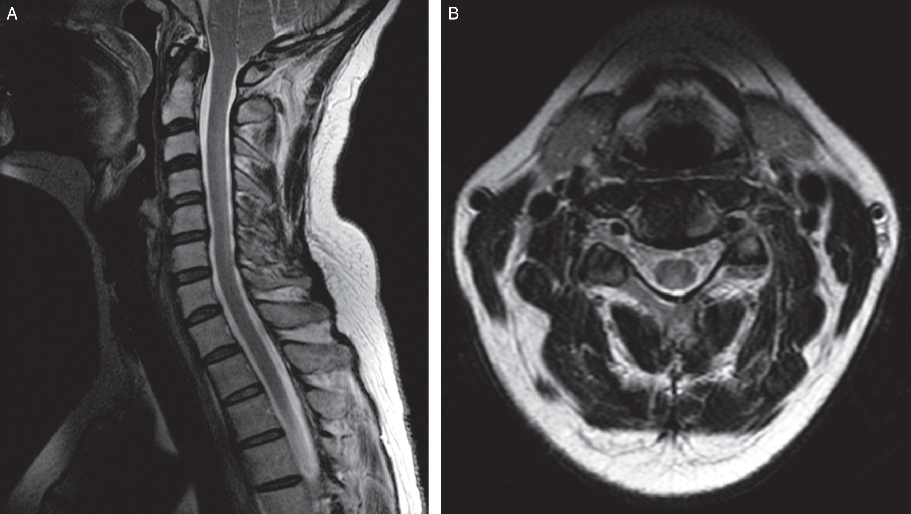
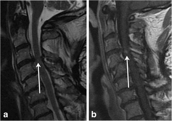
Diagnostic Approach to Intrinsic Abnormality of Spinal Cord Signal  Intensity | RadioGraphics
Diagnostic Approach to Intrinsic Abnormality of Spinal Cord Signal Intensity | RadioGraphics

Challenges in diagnosing spinal cord disease (Chapter 9) - Common Pitfalls  in Multiple Sclerosis and CNS Demyelinating Diseases
Challenges in diagnosing spinal cord disease (Chapter 9) - Common Pitfalls in Multiple Sclerosis and CNS Demyelinating Diseases

Diagnostic Approach to Intrinsic Abnormality of Spinal Cord Signal  Intensity | RadioGraphics
Diagnostic Approach to Intrinsic Abnormality of Spinal Cord Signal Intensity | RadioGraphics

Prognostic value of changes in spinal cord signal intensity on magnetic  resonance imaging in patients with cervical compressive myelopathy -  ScienceDirect
Prognostic value of changes in spinal cord signal intensity on magnetic resonance imaging in patients with cervical compressive myelopathy - ScienceDirect

Postoperative changes in spinal cord signal intensity in patients with spinal  cord injury without major bone injury: comparison between preoperative and  postoperative magnetic resonance images in: Journal of Neurosurgery: Spine  - Ahead
Postoperative changes in spinal cord signal intensity in patients with spinal cord injury without major bone injury: comparison between preoperative and postoperative magnetic resonance images in: Journal of Neurosurgery: Spine - Ahead

Peripheral Spinal Cord Hypointensity on T2-weighted MR Images: A Reliable  Imaging Sign of Venous Hypertensive Myelopathy | American Journal of  Neuroradiology
Peripheral Spinal Cord Hypointensity on T2-weighted MR Images: A Reliable Imaging Sign of Venous Hypertensive Myelopathy | American Journal of Neuroradiology
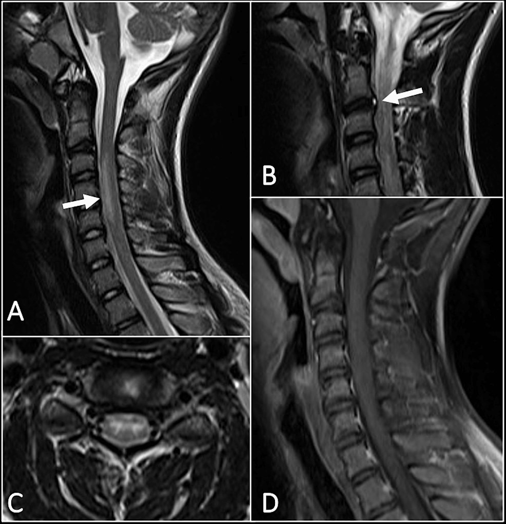
Cureus | Spinal Cord Infarct Due to Fibrocartilaginous Embolism in an  Adolescent Boy: A Case Report and Literature Review | Article
Cureus | Spinal Cord Infarct Due to Fibrocartilaginous Embolism in an Adolescent Boy: A Case Report and Literature Review | Article
A) Axial T2 weighted image of the spinal cord at the level of C1-2... |  Download Scientific Diagram
A) Axial T2 weighted image of the spinal cord at the level of C1-2... | Download Scientific Diagram

Location, length, and enhancement: systematic approach to differentiating  intramedullary spinal cord lesions | Insights into Imaging | Full Text
Location, length, and enhancement: systematic approach to differentiating intramedullary spinal cord lesions | Insights into Imaging | Full Text

MRI reveals abnormal high T2 cervical cord signal within the dorsal... |  Download Scientific Diagram
MRI reveals abnormal high T2 cervical cord signal within the dorsal... | Download Scientific Diagram
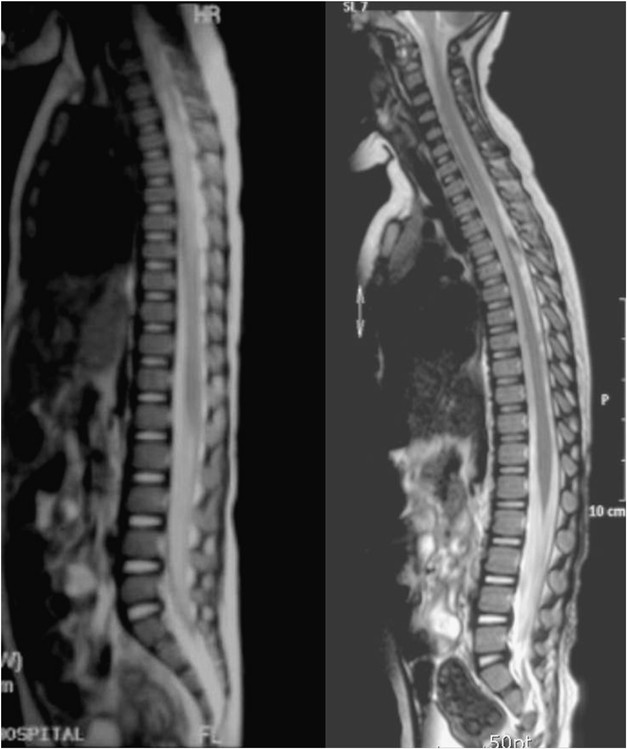
Progression of spinal cord atrophy by traumatic or inflammatory myelopathy  in the pediatric patients: case series | Spinal Cord
Progression of spinal cord atrophy by traumatic or inflammatory myelopathy in the pediatric patients: case series | Spinal Cord

Abnormal Spinal Cord Magnetic Resonance Signal: Approach to the  Differential Diagnosis - ScienceDirect
Abnormal Spinal Cord Magnetic Resonance Signal: Approach to the Differential Diagnosis - ScienceDirect

Spinal Cord Infarct | The Neurosurgical Atlas