Home

cable Anemone fish save apical cap heart Pirate cat Children

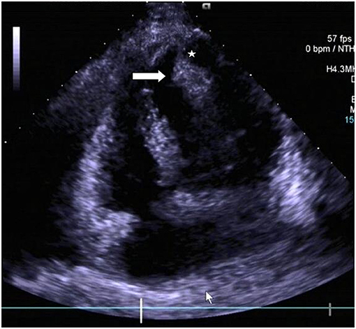
Apical 2-chamber view echocardiography, arrows show a thin-walled... |  Download Scientific Diagram
Apical 2-chamber view echocardiography, arrows show a thin-walled... | Download Scientific Diagram

Figure 8 from Recommendations for chamber quantification. | Semantic Scholar

A young man with chest pain | Tidsskrift for Den norske legeforening

Echocardiographic Evaluation of Coronary Artery Disease | Thoracic Key

Apical pleural cap | Radiology Reference Article | Radiopaedia.org

Apical Views/Coronary Arteries Flashcards | Quizlet

Pulmonary Apical Cap as a Potential Risk Factor for Pleuroparenchymal  Fibroelastosis - ScienceDirect
Pulmonary Apical Cap as a Potential Risk Factor for Pleuroparenchymal Fibroelastosis - ScienceDirect
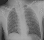
Apical pleural cap | Radiology Reference Article | Radiopaedia.org

Frontiers | Surgical Management for a Rare Pedunculated Left Ventricular  Apical Lipoma: A Case Report and Review of Literature
Frontiers | Surgical Management for a Rare Pedunculated Left Ventricular Apical Lipoma: A Case Report and Review of Literature

No Slide Title
No Slide Title

Apical Cap #radiology #xray #lungcancer - YouTube

Apical defect following Tendyne valve placement | SpringerLink

Apical left extrapleural cap: an early and important sign on chest  radiographs | Emergency Medicine Journal
Apical left extrapleural cap: an early and important sign on chest radiographs | Emergency Medicine Journal

Apical pleural cap | Radiology Reference Article | Radiopaedia.org

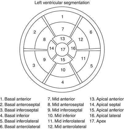
NephroPOCUS on X: "#POCUS Do you know what are the 17-segments of LV  assessed for regional wall motion abnormalities (RWMA)? As you may  recollect, #hemodialysis induces RMWAs in a significant proportion of
NephroPOCUS on X: "#POCUS Do you know what are the 17-segments of LV assessed for regional wall motion abnormalities (RWMA)? As you may recollect, #hemodialysis induces RMWAs in a significant proportion of

Conference Notes 4-18-2018 — ACMC EM

7: Myocardial segmentation into territories and segments. A:... | Download  Scientific Diagram
7: Myocardial segmentation into territories and segments. A:... | Download Scientific Diagram

Pleural Thickening: A List of Causes, Symptoms and Treatment

The segment division, endocardial boundary and the heart chambers are... |  Download Scientific Diagram
The segment division, endocardial boundary and the heart chambers are... | Download Scientific Diagram

The value of the left apical cap in the diagnosis of aortic rupture: a  prospective and retrospective study.
The value of the left apical cap in the diagnosis of aortic rupture: a prospective and retrospective study.

Left Atrial Appendage Closure Procedures | Johns Hopkins Medicine

Left Ventricular Rotation and Twist Assessed by Four‐Dimensional Speckle  Tracking Echocardiography in Healthy Subjects and Pathological Remodeling:  a Single Center Experience - Lilli - 2013 - Echocardiography - Wiley Online  Library
Left Ventricular Rotation and Twist Assessed by Four‐Dimensional Speckle Tracking Echocardiography in Healthy Subjects and Pathological Remodeling: a Single Center Experience - Lilli - 2013 - Echocardiography - Wiley Online Library

sufian_the_scribe on X: "Chest xray for thoracic aortic dissection Widened  mediastinum (56-63%), abnormal aortic contour (48%), aortic knuckle double  calcium sign >5mm (14%), pleural effusion (L>R), tracheal shift, left apical  cap, deviated
sufian_the_scribe on X: "Chest xray for thoracic aortic dissection Widened mediastinum (56-63%), abnormal aortic contour (48%), aortic knuckle double calcium sign >5mm (14%), pleural effusion (L>R), tracheal shift, left apical cap, deviated